Hormonal Control of Molting & Metamorphosis
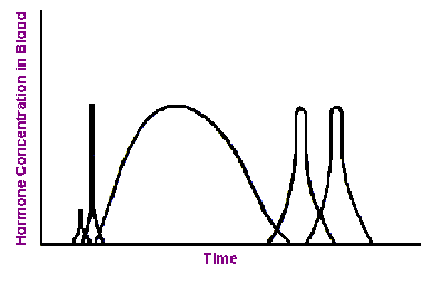
When an immature insect has grown sufficiently to require a larger exoskeleton, sensory input from the body activates certain neurosecretory cells in the brain. These neurons respond by secreting brain hormone which triggers the corpora cardiaca to release their store of prothoracicotropic hormone (PTTH) into the circulatory system. This sudden “pulse” of PTTH stimulates the prothoracic glands to secrete molting hormone (ecdysteroids).
 Molting hormone affects many cells throughout the body, but its principle function is to stimulate a series of physiological events (collectively known as apolysis) that lead to synthesis of a new exoskeleton. During this process, the new exoskeleton forms as a soft, wrinkled layer underneath the hard parts (exocuticle plus epicuticle) of the old exoskeleton. The duration of apolysis ranges from days to weeks, depending on the species and its characteristic growth rate. Once new exoskeleton has formed, the insect is ready to shed what’s left of its old exoskeleton. At this stage, the insect is said to be pharate, meaning that the body is covered by two layers of exoskeleton.
As long as ecdysteroid levels remain above a critical threshold in the hemolymph, other endocrine structures remain inactive (inhibited). But toward the end of apolysis, ecdysteroid concentration falls, and neurosecretory cells in the ventral ganglia begin secreting eclosion hormone. This hormone triggers ecdysis, the physical process of shedding the old exoskeleton. In addition, a rising concentration of eclosion hormone stimulates other neurosecretory cells in the ventral ganglia to secrete bursicon, a hormone that causes hardening and darkening of the integument (tanning) due to the formation of quinone cross-linkages in the exocuticle (sclerotization).
In immature insects, juvenile hormone is secreted by the corpora allata prior to each molt. This hormone inhibits the genes that promote development of adult characteristics (e.g. wings, reproductive organs, and external genitalia), causing the insect to remain “immature” (nymph or larva). The corpora allata become atrophied (shrink) during the last larval or nymphal instar and stop producing juvenile hormone. This releases inhibition on development of adult structures and causes the insect to molt into an adult (hemimetabolous) or a pupa (holometabolous).
At the approach of sexual maturity in the adult stage, brain neurosecretory cells release a brain hormone that “reactivates” the corpora allata, stimulating renewed production of juvenile hormone. In adult females, juvenile hormone stimulates production of yolk for the eggs. In adult males, it stimulates the accessory glands to produce proteins needed for seminal fluid and the case of the spermatophore. In the absence of normal juvenile hormone production, the adult remains sexually sterile.
Although the role of hormones in the physiology of molting was first described by V. B. Wigglesworth in the 1930’s, there is still much about the process that we do not fully understand. Insect endocrinology is currently an active area of research because it offers the potential for disrupting the life cycle of a pest without harm to the environment.

