The Head
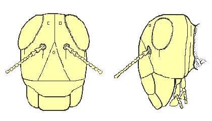
In most insects, the head capsule is a sturdy compartment that houses the brain, a mouth opening, mouthparts used for ingestion of food, and major sense organs (including antennae, compound eyes, and ocelli). Embryological evidence suggests that the first six body segments (three pre-oral and three post-oral) of a primitive worm-like ancestor may have fused to form the head capsule of most present-day insects.
The surface of the head is divided into regions (sclerites) by a pattern of shallow grooves (sutures). The uppermost sclerite (dorsal surface) of the head capsule is known as the vertex. A coronal suture usually runs along the midline of the vertex and splits into two frontal sutures as it extends downward across the front of the head capsule. The triangular sclerite that lies between these frontal sutures is called the frons. The epistomal suture is a deep groove that separates the base of the frons from the clypeus, a rectangular sclerite on the lower front margin of the head capsule.
The genae (“cheeks”) are lateral sclerites that lie behind the frontal sutures on each side of the head. Below each gena there may be another sclerite (the subgena), separated from the gena by a subgenal suture. A pair of compound eyes, sockets for two antennae, and one or more ocelli (simple eyes) also may be found on the front, top, or sides of an insect’s head.

Near the back of the head, an occipital suture circumscribes the head capsule at the posterior margin of the vertex and genae. This suture marks the location of an internal sclerotized ridge (apodeme) that strengthens this part of the head capsule. Just behind the occipital suture lie the occiput and postgenae, tiny sclerites that are probably remnants of the fifth primitive segment that fused to form the insect’s head. At the posterior-most margin of the head, a vestige of the sixth primitive segment is marked by a faint postoccipital suture and a thin, band-like sclerite (the postocciput) that adjoins the neck membrane.
The insect’s neck is known as the cervix. This is a membranous area that allows considerable freedom of movement for protraction and retraction of the insect’s head. The cervical membrane extends from the posterior portion of the postocciput to the prothorax, and it represents a transitional zone between the head and thorax. Small cervical sclerites serve as points of attachment for muscles that control head movements.
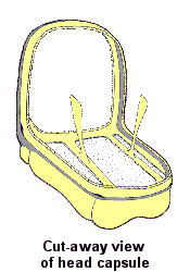
 Inside the head, a structure called the tentorium serves as an internal “truss” that reinforces the head capsule, cradles the brain, and provides a rigid origin for muscles of the mandibles and other mouthparts. The tentorium forms during development when pairs of apophyses (finger-like invaginations of exoskeleton) fuse internally to create a “bridge”. In most hemimetabolous insects, the tentorium is constructed from a pair of anterior arms and a pair of posterior arms (each “arm” represents a single apophysis). In some holometabolous insects, there are also a pair of dorsal arms that contribute to the structure. Externally, the location of each apophysis is often visible as a (anterior, posterior, or dorsal) “tentorial pit”.

