Digestive and Excretory Systems


An insect uses its digestive system to extract nutrients and other substances from the food it consumes.   Most of this food is ingested in the form of macromolecules and other complex substances (such as proteins, polysaccharides, fats, nucleic acids, etc.) which must be broken down by catabolic reactions into smaller molecules (i.e. amino acids, simple sugars, etc.) before being used by cells of the body for energy, growth, or reproduction.   This break-down process is known as digestion.

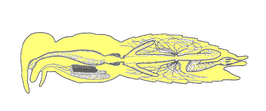
All insects have a complete digestive system.   This means that food processing occurs within a tube-like enclosure, the alimentary canal, running lengthwise through the body from mouth to anus.  Ingested food usually travels in only one direction.   This arrangement differs from an incomplete digestive system (found in certain lower invertebrates like hydra and flatworms) where a single opening to a pouch-like cavity serves as both mouth and anus.   Most biologists regard a complete digestive system as an evolutionary improvement over an incomplete digestive system because it permits functional specialization — different parts of the system may be specially adapted for various functions of food digestion, nutrient absorption, and waste excretion.   In most insects, the alimentary canal is subdivided into three functional regions:   foregut (stomodeum), midgut (mesenteron), and hindgut (proctodeum).

gut1a

In addition to the alimentary canal, insects also have paired salivary glands and salivary reservoirs.  These structures usually reside in the thorax (adjacent to the foregut).   Salivary ducts lead from the glands to the reservoirs and then forward, through the head, to an opening (the salivarium) behind the hypopharynx.   Movements of the mouthparts help mix saliva with food in the buccal cavity.

Stomodaeum

An insect’s mouth, located centrally at the base of the mouthparts, is a muscular valve (sphincter) that marks the “front” of the foregut.   Food in the buccal cavity is sucked through the mouth opening and into the pharynx by the action of cibarial muscles.   These muscles are located between the head capsule and the anterior wall of the pharynx.  When they contract, they create suction by enlarging the volume of the pharynx (like opening a bellows).   This “suction pump” mechanism is called the cibarial pump.   It is especially well-developed in insects with piercing/sucking mouthparts.   From the pharynx, food passes into the esophagus by means of peristalsis (rhythmic muscular contractions of the gut wall).  The esophagus is just a simple tube that connects the pharynx to the crop, a food-storage organ.

gut1a

Food remains in the crop until it can be processed through the remaining sections of the alimentary canal.   While in the crop, some digestion may occur as a result of salivary enzymes that were added in the buccal cavity and/or other enzymes regurgitated from the midgut.   In some insects, the crop opens posteriorly into a muscular proventriculus.   This organ contains tooth-like denticles that grind and pulverize food particles.   The proventriculus serves much the same function as a gizzard in birds.   The stomodeal valve, a sphincter muscle located just behind the proventriculus, regulates the flow of food from the stomodeum to the mesenteron.

In a developing embryo, the foregut arises as a simple invagination of the anterior body wall:   this means that all of its tissues and organs are derived from embryonic ectoderm.   In effect, the inside of the stomodeum is continuous with the outside of the insect’s body.   Since exoskeleton is secreted to protect the insect externally, it is not surprising to find that cells lining the foregut produce a similar structure (known as the intima) to protect themselves from abrasion by food particles.   The hard denticles inside the proventriculus are made from this same material.

Mesenteron

The midgut begins just past the stomodeal valve.   Near its anterior end, finger-like projections (usually from 2 to 10) diverge from the walls of the midgut.   These structures, the gastric caecae, provide extra surface area for secretion of enzymes or absorption of water (and other substances) from the alimentary canal.   The rest of the midgut is called the ventriculus — it is the primary site for enzymatic digestion of food and absorption of nutrients.   Digestive cells lining the walls of the ventriculus have microscopic projections (microvilli) that increase surface area for nutrient absorption.

gut1a

The midgut is derived from embryonic endoderm so it is not protected by an intima.   Instead, the midgut is lined with a semipermeable membrane secreted by a cluster of cells (the cardial epithelium) that lie just behind the stomodeal valve.   This peritrophic membrane consists of chitin fibrils embedded in a protein-carbohydrate matrix.   It protects the delicate digestive cells without inhibiting absorption of nutrient molecules.

The posterior end of the midgut is marked by another sphincter muscle, the pyloric valve.   It regulates the flow of material from the mesenteron into the proctodaeum.

Proctodaeum

The pyloric valve serves as a point of origin for dozens to hundreds of Malpighian tubules.   These long, spaghetti-like structures extend throughout most of the abdominal cavity where they serve as excretory organs, removing nitrogenous wastes (principally ammonium ions, NH4+) from the hemolymph.   The toxic NH4+ is quickly converted to urea and then to uric acid by a series of chemical reactions within the Malpighian tubules.   The uric acid, a semi-solid, accumulates inside each tubule and is eventually emptied into the hindgut for elimination as part of the fecal pellet.

gut1a

The rest of the hindgut plays a major role in homeostasis by regulating the absorption of water and salts from waste products in the alimentary canal.   In some insects, the hindgut is visibly subdivided into an ileum, a colon, and a rectum.   Efficient recovery of water is facilitated by six rectal pads that are embedded in the walls of the rectum.   These organs remove more than 90% of the water from a fecal pellet before it passes out of the body through the anus.

Embryonically, the hindgut develops as an invagination of the body wall (from ectodermal tissue). Just like the foregut, it is lined with a thin, protective layer of cuticle (intima) that is secreted by the endothelial cells of the gut wall. When an insect molts, it sheds and replaces the intima in both the foregut and the hindgut.